
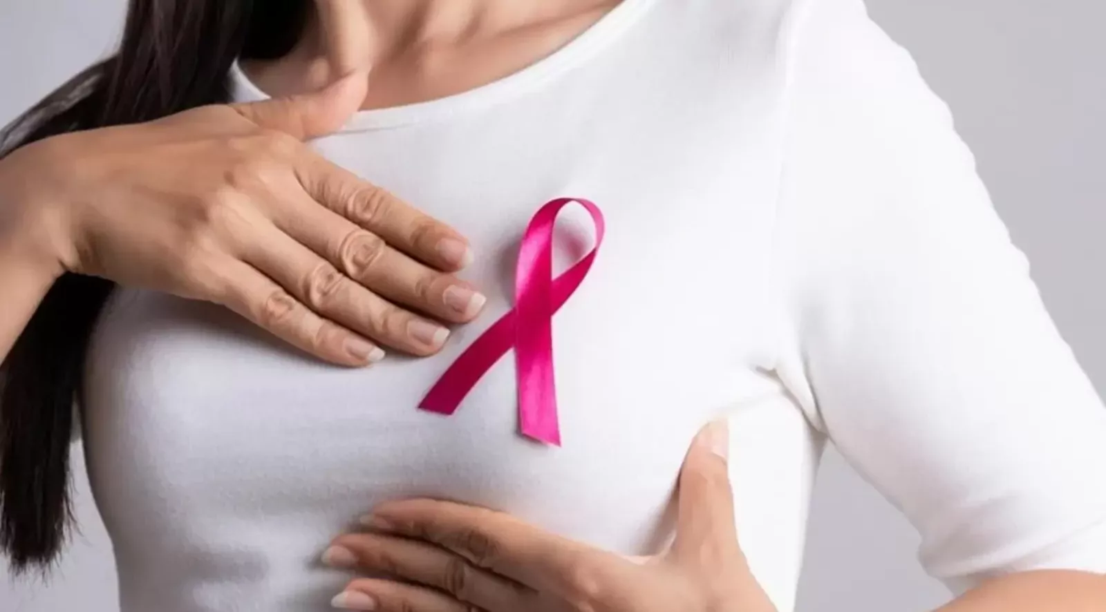
When you’re having a shower or fastening your underwear in front of the mirror and suddenly feel an unusual lump under your fingers, time seems to stand still. Your first reaction is fear, and hundreds of worrying questions spring to mind. At times like these, it’s important to take a deep breath and remind yourself of two facts: firstly, most breast lumps are benign, and secondly, even if it is a malignant condition, breast cancer is now one of the most well-understood and treatable forms of cancer. The main thing is not to panic, not to seek advice on forums, but to see a doctor straight away.
This article is intended for women who have noticed worrying changes or simply want to keep their health under control, as well as for loved ones looking for ways to support a family member. In modern oncology, understanding tumour biology and seeking specialist advice in good time It makes all the difference: as emphasised in the official clinical guidelines of the European Society for Medical Oncology (ESMO), it is early diagnosis, combined with improvements in treatment methods, that enables the disease to be managed and the highest rates of successful recovery to be achieved.
Why do tumours develop and how can you recognise the early signs?
Breast cancer develops from the epithelial cells of the ducts or lobules of the glandular tissue and, in its early stages, grows locally without causing any discomfort. It is precisely this insidious lack of symptoms that makes regular screening critically important, as pain is extremely rare in primary tumours, and the most common initial sign is a painless lump. Many women put off seeing a doctor precisely because «nothing hurts». As the tumour grows, it begins to affect the surrounding tissues, and visual changes then appear that cannot be ignored:
- Changes to the shape and skin of the breasts. Noticeable asymmetry, localised skin retraction over the lump, redness or swelling may occur. A particularly worrying symptom is the «orange peel» effect – thickening of the skin with enlarged pores, indicating a blockage of the lymphatic vessels.
- Changes to the nipple and areola. The nipple may become inverted, change colour, thicken or develop small sores and flaking similar to eczema. A warning sign is spontaneous discharge from one side – clear, yellowish or mixed with blood.
- Reaction of the lymph nodes. Enlarged and firm lymph nodes in the armpit or above the collarbone on the side of the affected breast are sometimes the first symptom, even if the lump in the breast itself is too small to be felt.
- Atypical and inflammatory manifestations. A rare but aggressive inflammatory form of breast cancer often does not form a distinct lump – the breast becomes hot to the touch, reddens, swells and becomes painful, which is often mistaken for mastitis. In women who are breastfeeding, a specific symptom has been described whereby the baby refuses the affected breast due to a change in the taste of the milk.
An important point for female patients: No single symptom in itself is a definitive diagnosis – fibroadenomas, cysts or mastopathy can masquerade as dangerous conditions. However, any new lump or change in the shape of the breast should be treated by a doctor as potentially malignant until an accurate diagnosis proves otherwise.

Modern diagnostics: from screening to a molecular profile of the tumour
Accurate diagnosis of breast cancer consists of two essential stages: imaging, which identifies a suspicious area, and a histological examination of the tissue, which establishes the final diagnosis. It is not possible to make a diagnosis based solely on a physical examination, an ultrasound scan or a mammogram – the final word always rests with the pathologist.
The choice of initial imaging method depends on the woman’s age and the density of her breast tissue. In younger patients, dense glandular tissue predominates; therefore, ultrasound (US) is the gold standard for them. After the age of 40–45, the tissue is gradually replaced by fatty tissue, and digital mammography comes to the fore, as it is capable of detecting microcalcifications and tumours measuring just a few millimetres long well before they can be felt by hand.
|
Survey method |
Who is it prescribed for, and when? |
What this allows the doctor to see |
|---|---|---|
|
Ultrasound scan of the breasts and lymph nodes |
Women under 40, pregnant women, breastfeeding women, and as a supplement to mammography |
Distinguishes fluid-filled cysts from solid masses, assesses blood flow within the lesion and the condition of the axillary lymph nodes |
|
Digital mammography |
A screening standard for women aged 40–45, as well as in cases where a tumour is suspected at any age |
Detects non-palpable tumours, asymmetry, abnormalities in tissue architecture and malignant microcalcifications |
|
Contrast-enhanced MRI of the breasts |
In cases of high genetic risk (BRCA mutations), complex diagnostic cases, or prior to planning surgery |
Determines the boundaries of the tumour, the presence of additional lesions and the extent of the disease with the greatest possible accuracy |
|
Trephine biopsy (thick-needle biopsy) |
If suspicious lesions classified as BI-RADS 4 or BI-RADS 5 are detected |
Provides a tissue section for histology, immunohistochemistry (IHC) and the determination of the biological subtype of cancer |
If, during a mammogram or ultrasound scan, the doctor identifies a suspicious area (BI-RADS category 4 or 5 on the international scale), an ultrasound-guided core needle biopsy is performed. Based on the results of histological and immunohistochemical analysis, specialists determine the tumour’s receptor status (sensitivity to oestrogen and progesterone), HER2 protein levels and the Ki-67 proliferation index. In Ukraine, it is possible to undergo a full cycle of expert diagnostics – from high-precision imaging to molecular genetic testing for BRCA1/2 mutations – and receive state-of-the-art treatment in accordance with international protocols at the specialised centre – the LISOD Israeli Oncology Hospital, where each clinical case is reviewed by a multidisciplinary panel of doctors.
Personalised treatment and realistic prognoses for breast cancer
Modern oncology has moved away from standardised treatment regimens: treatment strategies are always personalised. According to the updated NCCN guidelines, treatment combines local control (surgery, radiotherapy) with targeted systemic therapy.
The choice of treatment is based on the results of immunohistochemistry. For hormone-dependent cancer, the mainstay of treatment is endocrine therapy with CDK4/6 inhibitors; for HER2-positive cancer, targeted antibodies and conjugates; and for triple-negative cancer – combinations of chemotherapy with immunotherapy and PARP inhibitors (where BRCA1/2 mutations are present). At the same time, surgeons aim to perform minimally invasive, organ-preserving surgery or breast reconstruction.
The chances of recovery depend directly on the stage at which the disease is detected:
|
Stage ge[kbyb |
Prevalence of the process |
Estimated 5-year survival rate |
|---|---|---|
|
Stage I |
Tumour up to 2 cm; lymph nodes clear |
98–100% (full recovery) |
|
Stage II |
A tumour measuring 2–5 cm or isolated lymph nodes |
around 90 per cent |
|
Stage III |
A locally widespread process |
66–85% |
|
Stage IV* |
Presence of distant metastases |
22–25% (disease control) |
*Even at stage IV, innovative targeted and immunotherapy drugs make it possible to keep the disease at bay for years, whilst maintaining a high quality of life.
No online article or advice on a forum can replace a qualified face-to-face consultation with a breast cancer specialist. Any attempts at self-medication, a ‘wait-and-see’ approach or the use of dubious methods merely waste your most precious resource – time, which is of the utmost importance when it comes to cancer. If you have noticed any unusual changes in your breasts, are experiencing discomfort, or simply know that it is time for your next routine check-up – do not delay your visit to the hospital. Modern medicine has all the necessary tools for the early diagnosis and successful treatment of cancer, and seeing a doctor in good time is the simplest, yet most important, step towards preserving your health, peace of mind and longevity.